References:
- Kozier & Erb’s Fundamentals of Nursing: Concepts, Process, and Practice, 11th Edition, ISBN 9780135428733, by Audrey Berman, Shirlee J. Snyder, and Geralyn Frandsen (Ch. 29, pp. 604–614)
Assessing the thorax and lungs is frequently critical to assessing the client’s oxygenation status. Changes in the respiratory system can occur slowly (COPD) or quickly (pneumonia, embolus).
Chest Landmarks
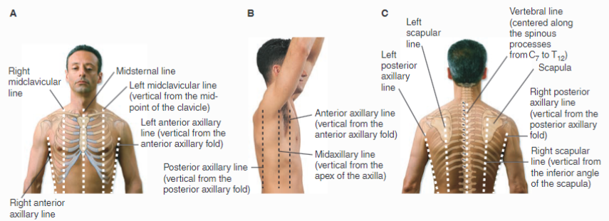
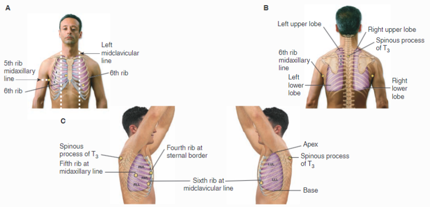
Chest landmarks are imperative for assessment. These include a series of imaginary lines on the chest wall and positions of ribs and spinous processes. These landmarks help to identify the position of underlying organs (e.g., lobes of the lung) and to record abnormal assessment findings. The chest wall is also divided into three regions: anterior, lateral, and posterior. The anterior chest wall includes the following items listed #1–3, the lateral chest walls include #3–5, and the posterior chest wall includes #6–8.
- Midsternal Line: A vertical line running through the center of the sternum.
- Midclavicular Lines: Vertical lines running through the midpoints of the clavicles (left and right).
- Anterior Axillary Lines: Vertical lines running down from the anterior axillary folds.
- Midaxillary Lines: Vertical lines running down from the apex of the axillae.
- Posterior Axillary Lines: Vertical lines running down from the posterior axillary folds.
- Vertebral Line: A vertical line centered through the spinous processes from C7 to T12
block of text