Reference:
- Dr. RPS Maternal and Newborn Care: A Comprehensive Guide and Source Book for Teaching and Learning, 2nd Edition, ISBN 9789719822653, by Rosalinda Parado Salustiano (Ch. 4, 64–79)
- Lecturer (V, delos Reyes)
Also Read
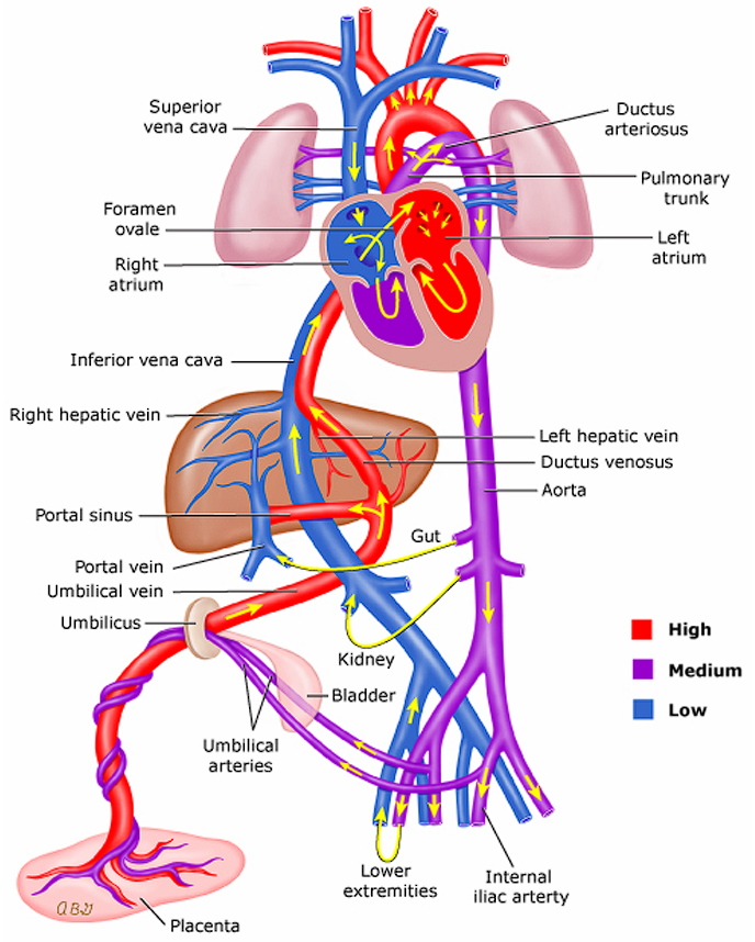
There are two external and three internal parts of fetal circulation that allows the fetus to oxygenate in-utero. These are the placenta, umbilical cord, ductus arteriosus, ductus venosus, and foramen ovale.
- Oxygenated blood from the placenta is sent through the umbilical vein through the ductus venosus and directly into the inferior vena cava. This bypasses the normal adult circulation through the liver. However, a minute amount of blood is still passed to the liver by the umbilical vein for nourishment. The inferior vena cava (oxygenated) and superior vena cava (deoxygenated) deposit blood into the right atrium.
- Blood coming in from the inferior vena cava will mostly pass through the foramen ovale into the left atrium, guided by the fetal eustachian valve. From the left atrium, blood is pumped to the left ventricle and through the aorta, which nourishes the brain.
- Blood coming in from the superior vena cava will mostly pass through the tricuspid valve into the right ventricle, through the pulmonary artery. The lungs are not yet functional, and so most of the blood will pass through the ductus arteriosus to the descending aorta.
- Deoxygenated blood is sent back to the placenta through the umbilical arteries from two arteries past the bifurcation of the descending aorta.
- The ductus (“ducts”; temporary connections between two vessels) shunts blood in fetal circulation. There are three shunts in a fetus:
- Ductus venosus: connects the umbilical vein and inferior vena cava, bypassing the immature liver.
- Ductus arteriosus: connects the pulmonary artery and aorta, allowing blood to shunt from right-to-left.
- Foramen ovale: connects the two atria, allowing blood to shunt left-to-right, bypassing the lungs.
- After clamping and cutting and at delivery, fetal circulation begins to adapt and become alike to adult circulation.
- The umbilical vessels (arteries and vein) will shrink and close.
- The baby’s lungs expand, which begin to receive more blood. This blood returns to the left atrium of the heart, increasing left-side pressure (and decreasing right-side pressure), closing the flap of the foramen ovale. It functionally closes in as early as 1 hour or about 1 to 3 days and anatomically closes within 2 to 3 months.
- Decreasing prostaglandin will also result in the closure of the ductus arteriosus as oxygen concentration increases, constricting the duct. It functionally closes in as early as 1 hour and about 1 to 3 days and anatomically closes within 2 to 3 months.
- The longest waiting time is 48 hours for the foramen ovale and ductus arteriosus to close before pathology is suspected. In assessment, these manifest as murmurs.
| Fetal Structure | Function | Time of Closure |
|---|---|---|
| Umbilical Vein | Carries oxygenated blood from the placenta to the inferior vena cava | At birth, then becomes the ligamentum teres |
| Umbilical Artery | Carries unoxygenated blood from the hypogastric arteries to the placenta | At birth, obliterated in 3 to 4 days, then becomes the umbilical ligaments |
| Ductus Venosus | Shunts blood from the umbilical vein to the inferior vena cava, bypassing the liver | At birth, then becomes the ligamentum venosum |
| Ductus Arteriosus | Shunts blood from the pulmonary artery to the descending aorta, bypassing the lungs | 1 hour to 3 days, then becomes the ligamentum arteriosum |
| Foramen Ovale | Shunts blood from the right atrium to the left atrium | 1 hour to 3 days, then becomes the fossa ovalis |
Tip!
Memorize the normal pathway of blood through the body and the heart. Visualization of how blood moves across each chamber and major vessel is very important in understanding the nature of congenital heart defects:
- IVC, SVC, and Coronary Vessels deposit blood into the Right Atrium
- Right Atrium, Tricuspid Valve, Right Ventricle
- Right Ventricle, Pulmonary Valve, Pulmonary Artery, Lungs
- Lungs, Pulmonary Veins, Left Atrium
- Left Atrium, Mitral/Bicuspid Valve, Left Ventricle
- Left Ventricle, Aortic Valve, Aorta