Reference:
- Brunner & Suddarth’s Textbook of Medical-Surgical Nursing, 15th Edition, ISBN 978-197-51-6103-3, by Janice L. Hinkle, Kerry H. Cheever, and Kristen J. Overbaugh (Ch. 59, pp. 1935-1963)
Anatomy and Physiology of the Ear
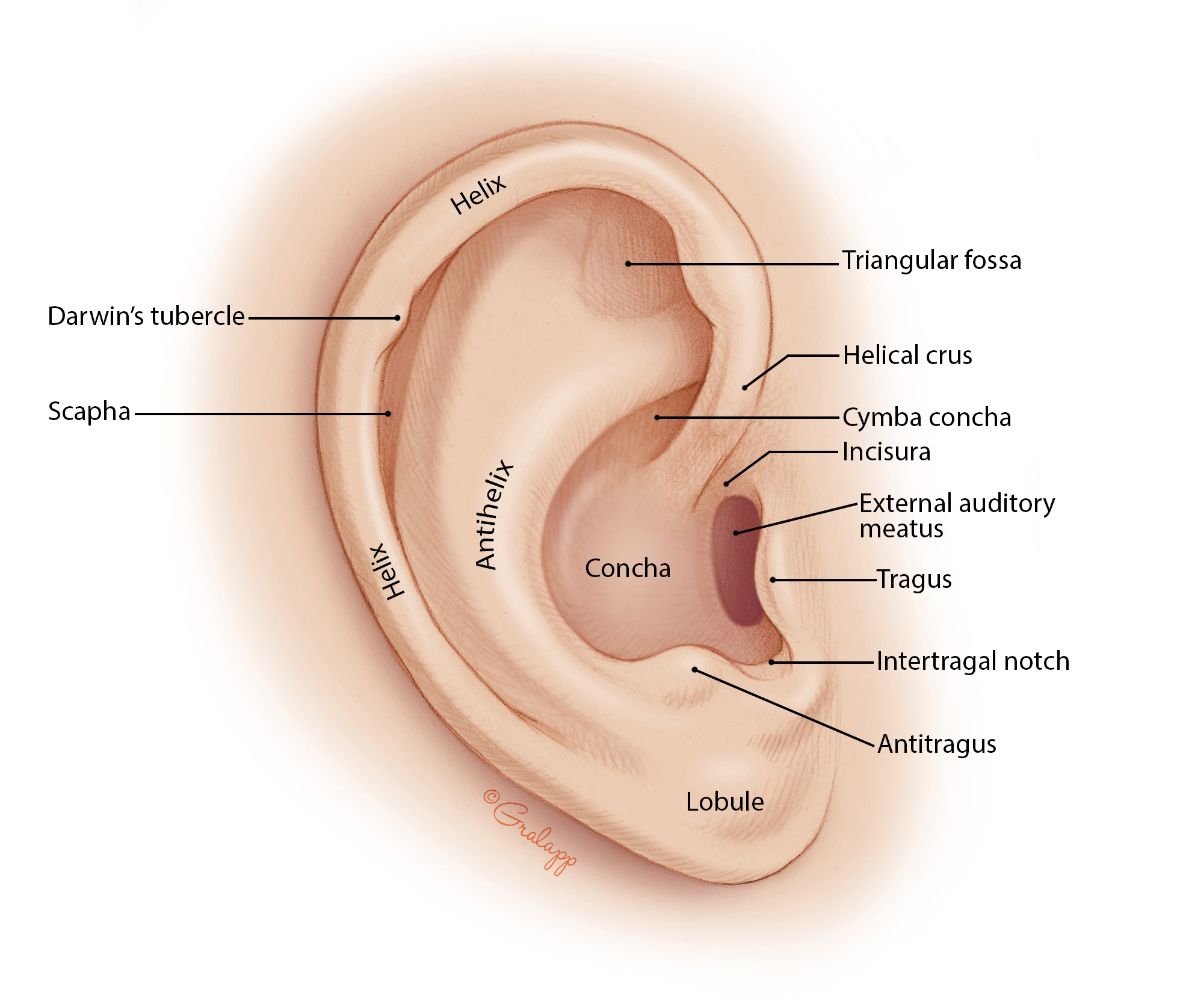
External Ear
The external ear consists of the auricle (pinna) and external auditory canal (ear canal). This region collects sound waves and directs them to the middle portion of the ear.
- Auricle, also known as the pinna, is a cartilaginous outer “funnel” that collects sound waves into the ear canal. This part is mainly made of cartilage (except for fat and subcutaneous tissue in the ear lobe).
- This is the part of the ear manipulated during otoscopy and otic medication administration
- External Auditory Canal, also known as the ear canal, is an approximately 2 to 3 cm long canal that ends at the tympanic membrane. This contains hair, sebaceous glands, and ceruminous glands (where ear wax, cerumen, comes from), which normally slowly clears the ear of debris and old cerumen. This may become impacted.
- Just anterior to the external auditory canal is the temporomandibular joint, whose head can be felt by placing a fingertip in the external auditory canal while the patient opens and closes their mouth.
Disorder
- An erythematous ear canal indicates otitis externa, inflammation of the ear canal.
- The ear canal is normally skin colored. If inflamed, it is erythematous.
- Aural tenderness occurs, where manipulation of the pinna, oracle, or canal causes pain.
- Treatment: analgesics (otic drops)
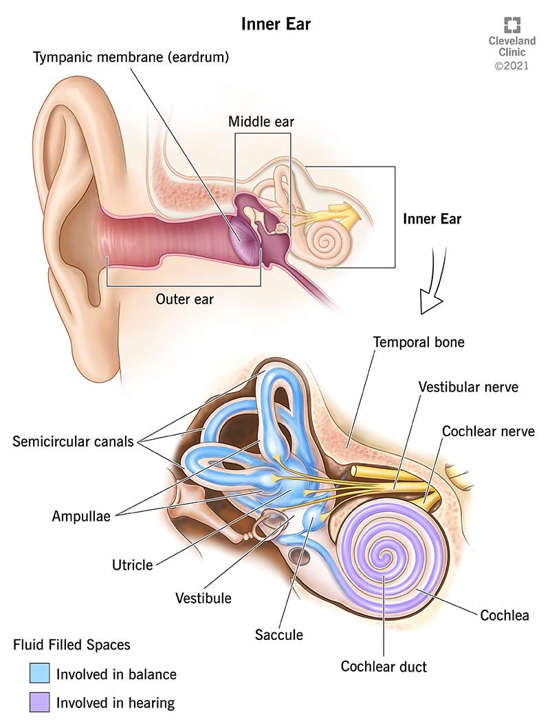
Middle Ear
The middle ear is an air-filled cavity, and includes the tympanic membrane/tympanum (eardrum), eustachian tube, the ossicles. The ossicles (malleus, incus, stapes) end at the oval window, which is what separates the middle ear from the inner ear.
- Tympanic Membrane: the structure separating the external ear from the middle ear. It is a thin, translucent, pearly gray membrane that protects the middle ear and conducts sound vibrations from the external canal to the ossicles.
- Ossicles: the three smallest bones of the body: malleus (hammer), incus (anvil), stapes (stirrups), connected by joints, muscles, and ligaments, conduct vibrations to the oval window, which separates the inner ear from the middle ear. Another fenestra, the round window, also on the border between the middle and inner ear, and acts as an exit point for the pressure waves.
Disorder
- Problems with the external or middle ear that results in hearing loss is conductive hearing loss.
- In otitis media, the tympanic membrane bulges and becomes erythematous (normally pearly grey).
- URTIs travelling through the eustachian tube can produce otitis media. In children, this tube is smaller, and more easily transfers infective agents.
Inner Ear
The inner ear is housed within the temporal bone. The organs for hearing (Cochlea), and balance (Semicircular Canals), as well as CN VII (Facial) and CN VIII (Vestibulocochlear). The cochlea and semicircular canals are housed in the Bony Labyrinth, which houses the Membranous Labyrinth. It is filled with endolymph and surrounded by perilymph. This fluid helps with the conversion of mechanical energy into neural impulses. The oval window connects to the vestibule, which is connected to both the semicircular canals responsible for balance, and the cochlea, responsible for hearing.
- Membranous Labyrinth: composed of the utricle, the saccule, the cochlear duct, the semicircular canals, and the Organ of Corti, all of which are surrounded by endolymph, a fluid.
- Semicircular Canals: there are three (posterior, superior, lateral) which lie at 90-degree angles to one-another. They contain sensory receptor organs that are arranged to detect rotational movement. These receptor end organs are stimulated by changes in the rate or direction of a person’s movement.
- The utricle and saccule are involved in linear movement. The utricle is horizontal-lying, and detects horizontal motion. The saccule is vertical, and detects motion along the sagittal plane; up, down, front, back.
- Organ of Corti: is the end organ for hearing. It is housed in the cochlea, a snail-shaped, bony tube about 3.5 cm long with two and a half spiral turns. It is located in the basilar membrane.
Disorder
- Labyrinthitis.
- Problems with the inner ear or labyrinth that results in hearing loss is sensorineural hearing loss. Often caused by (mn. LMP) labyrinthitis, meniere’s syndrome, and presbycusis**.
Physiology of Hearing
- The auricle (pinna) “gathers” sound waves, and redirects them to the external auditory canal and to the eardrum.
- The tympanum vibrates, sending vibrations across the bridge of ossicles, which connects with the oval window.
- The oval window vibrates as the stapes/stirrups strikes it, and vibrates the fluid within the vestibule.
- A round window lies further in the inner ear, and acts as a pressure valve; it bulges out when the inner ear pressure becomes elevated.
- The sensitive part of the inner ear, the Organ of Corti, functions as the body’s microphone with minuscule hairs that move with the endolymph’s waves. It is situated in the basilar membrane of one of the three components of the cochlea.
- The Vestibulocochlear Nerve, CN 8, facilitates hearing and equilibrium. The nerve fibers arise from the hearing and equilibrium apparatus of the inner ear, pass through the internal acoustic meatus, and enters the brainstem at the pons-medulla border. There are two divisions of this system: the cochlear (hearing) and vestibular (balance; equilibrium). CN 8 demonstrates purely sensory functions. The Facial Nerve, CN 7, innervates the stapedius muscle, which places tension on the bones of the middle ear. This tension regulates sound and protects the inner ear from damage due to loud sounds.
- The auditory cortex in the cerebrum then receive the impulses and decodes them as experienced sounds.
Functions of the Ear
- Hearing is the most basic function of the ear. Vibrations are interpreted as sound in the brain after being transmitted and transformed into nerve signals by the cochlea. This process may be done with air conduction (air-filled external and middle ear through vibration of the tympanic membrane and ossicles) or bone conduction, where the vibrations travel through bone and the inner ear, bypassing the tympanic membrane and ossicles. Air conduction is normally efficient at detecting vibrations.
- Sound Conduction and Transmission: sound enters through the (1) external auditory canal, and causes the (2) tympanic membrane to vibrate. The (3) ossicles, through lever action, transmit the sound as mechanical energy into the (4) oval window, transmitting it through the (5) inner ear fluids (perilymph, endolymph) to the (6) cochlea, stimulating (7) hair cells, converting the mechanical energy into electrical energy. These travel through the (8) vestibulocochlear nerve (CN VIII) to the (9) central nervous system, where it is interpreted.
- Sound conduction is impeded in a perforated tympanum, as this allows sound waves to apply pressure on the round window, which is normally an exit point for pressure waves.
- Balance is another important function. There are three semicircular canals in the inner ear. They are oriented at right angles to each other, and the resulting differences in the fluids of the canals helps the brain identify or detect the extent of movement and positioning of the head.
- Balance is achieved with the cooperation of muscles and joints in the body (proprioceptive system), the eyes (visual system), and the labyrinth (vestibular system). Each system informs the cerebellar system in the cerebral cortex about the body’s equilibrium for coordination and perception in the cerebral cortex. These, including the cardiovascular system that supplies the brain with blood, when damaged, all contribute to disturbance of balance.